
About this book
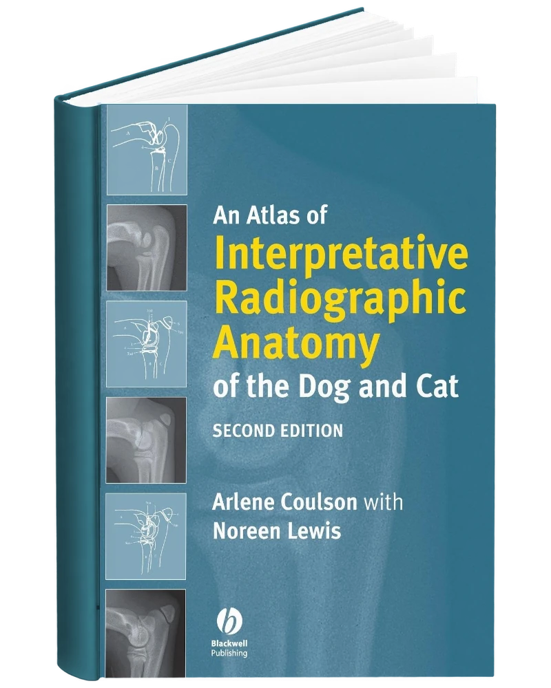
An Atlas of Interpretative Radiographic Anatomy of the Dog and Cat 2nd Edition PDF by Arlene Coulson and Noreen Lewis is offered here as a PDF eBook. This second edition is a detailed reference to normal radiographic anatomy in dogs and cats, created to help readers distinguish expected anatomy and normal variation from disease. High-quality radiographs are paired with detailed line drawings that identify important structures and make difficult regions easier to interpret. The atlas covers the skeletal system, including limbs, skull, vertebral column, ribs and sternum, as well as soft-tissue anatomy of the pharynx, larynx, thorax and abdomen. It also addresses normal variations associated with age, breed and development. The visual format makes it particularly valuable for veterinary students and practitioners who need a dependable baseline before interpreting abnormal diagnostic images.
Designed for veterinary students, small-animal practitioners, and veterinary imaging trainees, this edition provides a structured resource for understanding how anatomical structures appear on canine and feline radiographs.
This listing is for the 2nd edition of An Atlas of Interpretative Radiographic Anatomy of the Dog and Cat. Check the title, edition, authors/editors, and other product information before ordering if you need the book for a particular course, syllabus, examination, or professional reference.
About This Canine and Feline Radiographic Atlas
This atlas is centered on the normal radiographic anatomy that underlies interpretation of small-animal images. Arlene Coulson and Noreen Lewis organize the material into plain and contrast radiography, with dog and cat sections covering skeletal and soft-tissue structures. Interpretative line drawings help readers relate radiographic shadows to the anatomy responsible for them. The atlas also considers variation between breeds and in juvenile animals, along with appearances that can create interpretative pitfalls. The second edition adds figures and revised reference organization. Its focus makes it particularly useful for learning what belongs within the normal range before considering whether an image shows an abnormal finding.
What the Radiographic Atlas Covers
The 2nd edition covers the following areas:
- Canine skeletal anatomy: Normal structures shown through plain radiographic projections.
- Canine soft tissues: Radiographic appearances of soft-tissue anatomical areas.
- Feline skeletal anatomy: Species-specific skeletal reference images.
- Feline soft tissues: Normal soft-tissue appearances in cats.
- Contrast radiography: Contrast studies for canine and feline anatomical interpretation.
- Normal variation: Differences associated with age and breed.
- Interpretative pitfalls: Appearances that may complicate recognition of normal anatomy.
- Line and schematic drawings: Visual explanations linking image shadows with underlying structures.
Using This Atlas to Interpret Small-Animal Images
Normal anatomy is the comparison point for interpreting a radiograph. This atlas is useful because it brings that comparison into a visual format, pairing radiographic appearances with drawings that explain the structures behind them. A student can use it to relate a projection to an anatomical region, while a practitioner can consult it when an unfamiliar normal variant makes an image difficult to interpret. The separate canine and feline material also helps readers avoid assuming that the same appearance applies across species. Its practical value is in supporting careful anatomical recognition and image discussion, including the distinction between an expected variation and an appearance that warrants further investigation.
Who uses this book
- Veterinary Students Learning Diagnostic Imaging
- Small Animal Veterinary Practitioners
- Veterinary Radiology Residents and Trainees
- Veterinary Interns and Postgraduate Trainees
- Veterinary Surgeons Reviewing Normal Radiographic Anatomy
- Emergency and Critical Care Veterinarians Interpreting Radiographs
- Veterinary Technicians Studying Diagnostic Imaging
- Veterinary Diagnostic Imaging Educators
- Clinicians Comparing Normal Canine and Feline Anatomical Variations
Also suitable for other small-animal professionals who need an image-based reference to normal canine and feline radiographic anatomy before evaluating pathology.
Key Topics Covered
- Canine radiographic anatomy
- Feline radiographic anatomy
- Plain radiography
- Contrast radiography
- Skeletal projections
- Soft-tissue radiographic anatomy
- Juvenile anatomical variation
- Breed-related variation
- Radiographic shadows
- Interpretative line drawings
- Schematic anatomy
- Radiographic pitfalls
PDF Edition Features
- Searchable reading: Quickly locate names, concepts, conditions, terminology, and other material with a compatible PDF reader.
- Highlighting and notes: Use annotation, bookmarking, and highlighting features supported by your chosen PDF application.
- Flexible access: Read the eBook on compatible computers, tablets, and mobile devices.
- Edition identification: Check the edition and product information before ordering to ensure it matches your requirements.
- Instant access: After successful payment, the download link is available through your order and sent to the email address used at checkout.



Reviews
There are no reviews yet.