
About this book
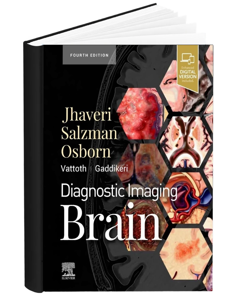
Diagnostic Imaging Brain 4th Edition PDF by Miral D Jhaveri, Karen L Salzman, Anne G Osborn, Surjith Vattoth and Santhosh Gaddikeri is offered here as a PDF eBook. Diagnostic Imaging: Brain provides a visual framework for studying common and uncommon neuroimaging diagnoses. The fourth edition approaches brain and central nervous system (CNS) conditions through both anatomy-based and pathology-based organization. Annotated images help readers connect location and appearance with the relevant disease discussion. Concise, templated chapters make it easier to compare findings across diagnostic entities. The edition includes topics such as autoimmune encephalitis, critical illness-associated microbleeds and selected congenital and infectious conditions. It also discusses brain tumor classification as understood in this edition, including the role of histological and molecular features. Coverage of magnetic resonance imaging (MRI) and computed tomography (CT) developments adds technical context to the diagnostic material. The book supports detailed neuroradiology learning and focused reference reading around an unfamiliar imaging pattern.
Brain Imaging and Neurological Differentials in the 4th Edition
Diagnostic Imaging: Brain connects neuroradiological findings with the neurological conditions that produce them. The fourth edition supports anatomical and pattern-based study of intracranial disease through imaging examples and focused diagnostic discussions.
Subjects Covered
- Cerebrovascular disease
- Brain tumors
- Infection
- Developmental abnormalities
- White matter disease
Using Diagnostic Imaging Brain for Study and Reference
For a case-based revision session, localize the finding before reading the relevant condition. Record the imaging observations separately from the proposed explanation, and compare a related entity to see which questions remain unresolved. That discipline helps a discussion move beyond pattern recognition alone. A study notebook can also link neurological terminology with the structures shown on imaging. The book provides educational examples and background; an actual examination must be interpreted by qualified professionals using the complete imaging study, clinical context and current standards.
Compare an intracranial imaging pattern with its anatomical distribution, then review the competing diagnostic explanations. Use the text alongside the reading list or training materials for your course or specialty, and check that the edition corresponds to your requirements.
Who uses this book
- Radiology Residents
- Neuroradiology Fellows
- Practicing Neuroradiologists
- General Radiologists
- Neurology Trainees Studying Neuroimaging
- Neurosurgery Trainees Studying Brain Imaging
- Radiology Educators
- Medical Students Undertaking Advanced Radiology Study
Also suitable for other neurological clinicians seeking specialist imaging background, with diagnostic examples interpreted alongside the full examination and clinical context.
PDF Reading Features
- Searchable PDF: Locate terminology and topics with text search in a compatible PDF reader.
- Notes and highlighting: Annotation and bookmarking depend on the features of your chosen reading application.
- Device access: Read the file on a compatible computer, tablet or smartphone.
- Instant access: After successful payment, the file link appears in your order details and is sent to the checkout email address.
This reference is for education and professional study. Clinical decisions require current guidance and qualified professional judgment. Publisher platforms, access codes, videos and other separately hosted resources are not included in this PDF listing.



Reviews
There are no reviews yet.